

X-ray equipment is one of the most important when it comes to diagnostic video imaging medical equipment. This is because the device was the forerunner of all other technologies in this area, such as tomography, mammography, and magnetic resonance devices.
But what was already good got even better. Today, radiological equipment is available in many types, sizes, and prices. So, if you are thinking of purchasing an RX machine or want to learn more about it, keep reading this article.
As you read, you will know who invented the X-ray, how it works, the main exams performed, and the differentials of digital radiography compared to the conventional X-ray machine.
In addition to the history and technology behind the equipment, I will talk about reporting options available for your healthcare facility. Shall we go ahead?
What Is An X-Ray Machine For?
X-ray equipment is used to take x-rays, which are like photographs of the inside of the body. Through these images, it is possible to observe anatomical structures, such as bones, organs, and blood vessels, without the need for surgery.
As it is cheap and non-invasive, nor does it cause pain, the exam is one of the most requested physicians worldwide. Thus, the X-ray device has been helping health professionals for decades, facilitating the diagnosis of pathologies located in different parts of the body.
Tumors, fractures, blocked blood vessels and even cavities are some of the ailments that can be identified utilizing an X-ray.
Who Invented The X-Ray?
Credit goes to German physicist Wilhelm Conrad Roentgen. In 1895, he discovered X-rays and their use to record internal images of the human body. Afterward, he shared his observations through the article “On a New Species of Rays.”
Magnetic Resonance Reports
The first radiograph dates from December 22 of that year, when the physicist placed the hand of his wife, Anna Bertha Roentgen, on the chassis with which he had been carrying out experiments. The procedure lasted about 15 minutes and made history.
In developing the film, Roentgen confirmed his conclusions by observing an image of his wife’s hand. In which bones and soft tissue could be visualized. In 1901, the specialist received the Nobel Prize in Physics for his discovery.
What Are The Types Of X-Ray Equipment?
The devices used in radiographs have undergone considerable evolution since the discoveries of Roentgen. In addition to conventional equipment, there are digital, mobile, and those used in fluoroscopy with analog technology. Let’s meet them?
Conventional X-Ray Equipment
The device is usually fixed in the room used for X-rays. It consists of a horizontal table that moves in all directions to focus on the body part to be examined. In the upper area is the X-ray tube, which has the components responsible for producing radiation, an opening through which the X-ray beam and collimators exit.
After capturing the images, this equipment records them on a film composed of silver halide salts. Therefore, to visualize the photos taken during the radiography, it is necessary to develop this film.
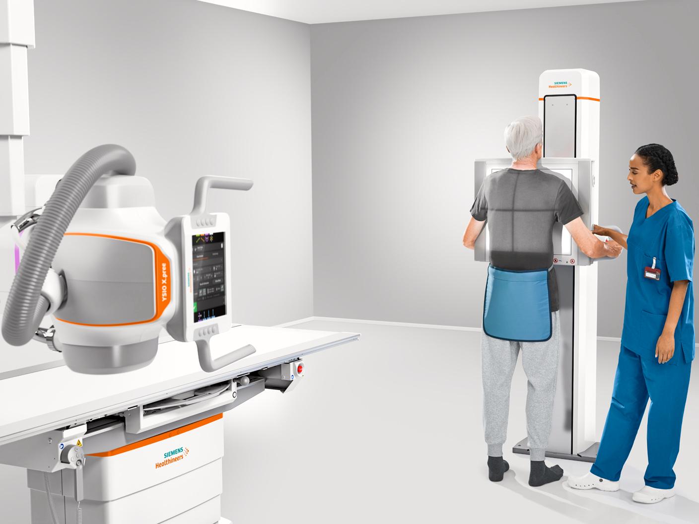
Digital X-Ray Equipment
The main difference between the conventional and the digital device is capturing and forming the images. As mentioned above, conventional equipment uses a film to record the radiography information.
Digital, on the other hand, captures the data through a radiation-sensitive plate and then forms the pixel images in direct or indirect connection with the computer. Thus, Digital technology also makes it possible to obtain clear images with reduced patient exposure time to X-rays.